Normal Female Pelvic Ultrasound Images - Consider Ultrasound First For Imaging The Female Pelvis American Journal Of Obstetrics Gynecology : See more ideas about ultrasound, sonography, ultrasound sonography.
Normal Female Pelvic Ultrasound Images - Consider Ultrasound First For Imaging The Female Pelvis American Journal Of Obstetrics Gynecology : See more ideas about ultrasound, sonography, ultrasound sonography.. A transabdominal (ta) evaluation and a transvaginal (tv) / endovaginal (ev) evaluation. An ultrasound review of pelvic pathology judi m bender md january 20, 2016. If not, one will be performed at first visit, then subsequent exams will be limited to areas of interest. The patient must have had a documented complete pelvic ultrasound within the last 6 months; A transabdominal (ta) evaluation and a transvaginal (tv) / endovaginal (ev) evaluation.
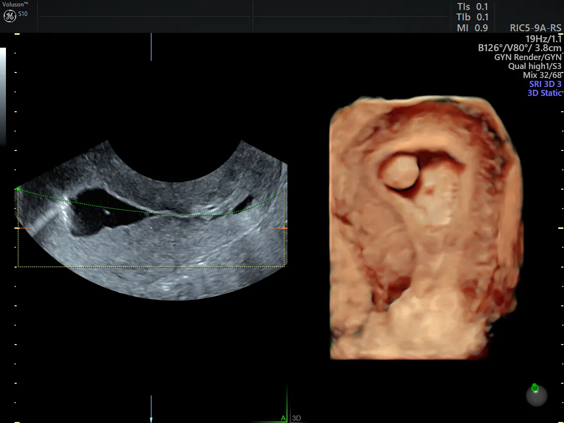
Whether the female pelvis is imaged because of a suspicion of underlying gynecologic disease or as a screening examination in the asymptomatic patient, those who interpret the images should be familiar with the range of normal appearances in these organs to avoid misinterpreting expected physiologic changes as pathologic conditions and to spare. A pelvic ultrasound allows quick visualization of the female pelvic organs and structures including the uterus, cervix, vagina, fallopian tubes and ovaries. The true pelvis is bounded anteriorly by the pubis and pubic rami, posteriorly by the sacrum and coccyx, laterally by the fused ilium and ischium, and inferiorly by the muscles of the pelvic floor. Multiple sonographic images of the pelvis were assessed for gray scale appearance and color doppler flow. The endometrial stripe measures __ cm in diameter and is normal in echogenicity.
How To Perform A Gynaecological Ultrasound In The Paediatric Or Adolescent Patient Deslandes 2020 Australasian Journal Of Ultrasound In Medicine Wiley Online Library from onlinelibrary.wiley.com A computer within the ultrasound machine turns this information into images that are displayed on a black and white screen. If a male sonographer is doing the scan, there will need to be a female chaperone present for the transvaginal or translabial portion of the exam. Langer, md • edward r. Pelvic imaging through the life cycle 1575 jill e. An ultrasound review of pelvic pathology judi m bender md january 20, 2016. However, it is considered more invasive than the transabdominal approach. Pathology not covered •pelvic pathology related to pregnancy, congenital or childhood abnormalities or normal variants •pelvic pathology related to non gynecological problems. Premenopausal women •simple cysts •1.<3cm=normal finding •2.
The test can be done in two ways:
If you want to learn more, i invite you to keep reading. If the transducer is moved over the abdomen it is known as a. See more ideas about ultrasound, sonography, uterus. Ultrasound of the pelvis protocol. In many ultrasound laboratories, the standard protocol for ultrasound examination of the female pelvis begins with tas using the filled urinary bladder as the acoustic window, followed by tvs after emptying the bladder and placing the patient in the lithotomy position. A pelvic ultrasound allows quick visualization of the female pelvic organs and structures including the uterus, cervix, vagina, fallopian tubes and ovaries. If a male sonographer is doing the scan, there will need to be a female chaperone present for the transvaginal or translabial portion of the exam. Premenopausal women •simple cysts •1.<3cm=normal finding •2. The sound waves create a picture on a video monitor. An ultrasound review of pelvic pathology judi m bender md january 20, 2016. The exam normally involves two components: If not, one will be performed at first visit, then subsequent exams will be limited to areas of interest. Sonography plays the primary role in imaging of the female pelvis.
If not, one will be performed at first visit, then subsequent exams will be limited to areas of interest. Ultrasound of the pelvis protocol. In many ultrasound laboratories, the standard protocol for ultrasound examination of the female pelvis begins with tas using the filled urinary bladder as the acoustic window, followed by tvs after emptying the bladder and placing the patient in the lithotomy position. Color doppler ultrasound image on right shows normal vascularity of the pelvic kidneys. There are two ways to create the pictures:
Common Perimenopause Symptoms Ultrasound Diagnosis Empowered Women S Health from images.takeshape.io Female pelvis ultrasound protocol the patient should be scanned either trans abdominally (ta) with a full bladder or trans vaginally (tv). Color doppler ultrasound image on right shows normal vascularity of the pelvic kidneys. The sound waves create a picture on a video monitor. See more ideas about ultrasound, sonography, uterus. The uterus measures __ x __ x __ cm in length, width and height respectively. Pelvic imaging through the life cycle 1575 jill e. Post surgical complications eg abscess, oedema. It allows your doctor to see your bladder, cervix, uterus, fallopian tubes, and ovaries.
Oliver, md, phd • anna s.
However, it is considered more invasive than the transabdominal approach. Über 7 millionen englischsprachige bücher. See more ideas about ultrasound, sonography, uterus. The sound waves create a picture on a video monitor. Langer, md • edward r. In the past, the cost of the mri was significantly higher than ultrasound and that often prevented people from using pelvic mri more often. Another ultrasound image (right) shows the bilateral pelvic kidneys adjacent to the ovaries and posterior to the urinary bladder. Guidance of injections, aspiration or biopsy. Premenopausal women •simple cysts •1.<3cm=normal finding •2. Pelvic ultrasound is usually the initial modality for imaging gynecologic pathology, including acute pelvic pain and chronic pelvic pain. Female pelvis ultrasound protocol the patient should be scanned either trans abdominally (ta) with a full bladder or trans vaginally (tv). The resolution of the ultrasound machines is significantly lower than the pelvic mri. Ultrasound of the pelvis protocol.
Ultrasound of the pelvis protocol. See more ideas about ultrasound, sonography, ultrasound sonography. Langer, md • edward r. If you want to learn more, i invite you to keep reading. In the past, the cost of the mri was significantly higher than ultrasound and that often prevented people from using pelvic mri more often.
Multiple sonographic images of the pelvis were assessed for gray scale appearance and color doppler flow.
Post surgical complications eg abscess, oedema. With a straight shot, the image produced will show the left side of your body on the right side of the image (just like a photograph). Fed up with deciphering jargon, dr attiya khan asked consultant gynaecologist mr rehan khan for a plain language guide to understanding pelvic ultrasounds. Written reports and ultrasound images/video clips that contain. If the transducer is moved over the abdomen it is known as a. By dr attiya khan and mr rehan khan. Primary indications for female pelvic us examination are pelvic pain, abnormal vaginal bleeding, and suspicion of pelvic mass. In the past, the cost of the mri was significantly higher than ultrasound and that often prevented people from using pelvic mri more often. Ultrasound of the female pelvis is important for diagnostic accuracy. See more ideas about ultrasound, sonography, uterus. Overview pelvic sonography is the imaging modality of choice for evaluating the female pelvis. Complete pelvic ultrasound (upeltv) this is a complete pelvic ultrasound exam, including transabdominal and transvaginal. The patient must have had a documented complete pelvic ultrasound within the last 6 months;
Sonography plays the primary role in imaging of the female pelvis pelvic ultrasound female. In the absence of masses in the nongravid patient, the uterus, ovaries, and adnexa are situated in the true pelvis.


0 Komentar